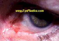
The eyelid is one of the most common sites for skin tumors on the body, both benign and malignant. Because the eyelid protects the eye, even small tumors in this location have functional and aesthetic significance. Oculoplastic surgeons evaluate, biopsy, remove, and reconstruct eyelid tumors as a single-surgeon team — a key advantage when Mohs surgery and oculoplastic reconstruction must be coordinated on the same day.
The first step with any new eyelid lesion is accurate diagnosis. Clinical features, growth rate, and patient history guide whether a lesion needs observation, office biopsy, or excision. When cancer is suspected, margin-controlled excision (Mohs micrographic surgery or frozen-section control) is standard of care before reconstruction.
Malignant Eyelid Lesions
Malignant eyelid tumors are predominantly epithelial in origin. The eyelid receives intense cumulative UV exposure — particularly the lower lid and medial canthus — and harbors both sebaceous glands and melanocytes, giving rise to a broad range of malignancies. The four most clinically important are basal cell carcinoma (most common), sebaceous carcinoma, squamous cell carcinoma, and melanoma.
General principles of management:
All suspicious lesions are biopsied before definitive excision — tissue diagnosis drives surgical planning
Margin-controlled excision (Mohs or frozen section) achieves the lowest recurrence rates for periocular basal and squamous cell carcinoma; melanoma is often excised with wider margins and permanent-section (delayed) margin control, since frozen sections are unreliable for melanocytic lesions, and sebaceous carcinoma may require conjunctival map biopsies to detect pagetoid spread
Reconstruction is planned at the time of excision; defect size and location determine the technique (direct closure, advancement flap, rotation flap, full-thickness graft, or staged Hughes tarsoconjunctival flap)
Sentinel lymph node biopsy is considered for high-risk squamous cell carcinoma, sebaceous carcinoma, melanoma, and Merkel cell carcinoma
Red flags for malignancy: loss of lashes (madarosis), induration or firmness beyond the visible lesion, irregular or pearly border, telangiectatic vessels, ulceration, recurrence after treatment, or any lesion that bleeds spontaneously.
When Is an Eyelid Bump Worrying?
Most eyelid bumps are harmless — a stye, a chalazion, a skin tag, or a xanthelasma. But certain features suggest a skin cancer and warrant prompt evaluation and biopsy:
A lesion that bleeds, ulcerates, or crusts and will not heal
Loss of eyelashes (madarosis) in the area of the bump
Irregular borders, mixed or changing color, or pearly rolled edges
Distortion of the normal lid margin or architecture
A “chalazion” that keeps recurring in the same spot — a red flag for sebaceous carcinoma
Benign vs. Malignant Lesions
Benign growths — papillomas, cysts, syringomas, and other benign lesions — are common and usually removed for comfort or appearance; removed tissue is routinely sent for pathologic examination to confirm the lesion is truly benign. Malignant eyelid tumors, in rough order of frequency, are:
Basal cell carcinoma — by far the most common, slow-growing, and rarely metastasizes — but if neglected (especially at the medial canthus) it can invade deeply into surrounding tissue and the orbit, so early treatment matters.
Squamous cell carcinoma — less common, more aggressive.
Sebaceous gland carcinoma — rare, aggressive, notorious for masquerading as a recurrent chalazion or chronic blepharitis.
Diagnosis begins with a biopsy. Confirmed eyelid cancers are usually excised with margin control — often Mohs micrographic surgery, which removes the tumor layer by layer while checking margins under the microscope, sparing as much healthy lid as possible. Because the eyelid protects the eye, reconstruction by an oculoplastic surgeon is as important as the cancer removal: the repair must restore a smooth lid margin, a stable tear film, and full closure to keep the eye safe.
Prevention & Choosing a Surgeon
Lifelong sun protection — sunglasses and sunscreen — lowers risk, and any suspicious or non-healing lid lesion should be checked early. Eyelid tumor removal and reconstruction are best handled by an ASOPRS fellowship-trained oculoplastic surgeon, who can both clear the cancer and rebuild the lid.
Frequently Asked Questions
What are the most common eyelid skin cancers?
Basal cell carcinoma (BCC) is the most common eyelid malignancy, accounting for roughly 90% of cases. Squamous cell carcinoma (SCC) is second. Sebaceous gland carcinoma, though rare, is especially dangerous because it mimics inflammatory conditions (masquerade syndrome). Melanoma can also occur on the eyelid skin.
How is eyelid skin cancer removed?
Eyelid skin tumors are excised with margin control — either by Mohs micrographic surgery (performed by dermatology) or by frozen-section control intraoperatively. After tumor clearance, the oculoplastic surgeon performs reconstruction of the eyelid, which may involve flaps, grafts, or complex multi-layer techniques to restore both function and appearance.
Can eyelid skin cancer spread to the eye?
Basal cell carcinoma rarely metastasizes but can invade deeply into the orbit if neglected. Sebaceous gland carcinoma has a significant risk of pagetoid spread (spreading along the conjunctival surface) and regional lymph node metastasis. Melanoma has the highest metastatic potential of any eyelid skin cancer.
What should I expect during my consultation for eyelid skin tumor evaluation?
During your consultation, Dr. Fountain will perform a thorough examination of the eyelid lesion, including its size, location, and characteristics. They may use specialized imaging or take a biopsy to confirm the diagnosis and determine if the tumor is benign or malignant. Dr. Fountain will discuss treatment options, show you before-and-after examples, and explain what to expect during surgery and recovery.
What happens after eyelid skin tumor removal regarding scarring and appearance?
Dr. Fountain will use advanced reconstruction techniques to minimize scarring and preserve normal eyelid function and appearance. The final appearance depends on the tumor's size and location, but fellowship-trained oculoplastic surgeons are specially trained to achieve cosmetically pleasing results while ensuring complete tumor removal. Most scars fade significantly over 6-12 months as they mature.
What are the potential risks and complications of eyelid tumor surgery?
While eyelid tumor surgery is generally safe, potential risks include infection, bleeding, and temporary swelling or bruising. Rarely, complications may affect eyelid function, such as difficulty closing the eye or changes in eyelid position, though experienced oculoplastic surgeons take specific precautions to prevent these issues. Dr. Fountain will discuss all risks during your consultation and explain how they minimize these complications.
How should I care for my eyelid after tumor removal surgery?
After surgery, you'll need to keep the area clean and dry, apply prescribed ointments, and avoid strenuous activity for 1-2 weeks. Dr. Fountain will provide detailed post-operative instructions and schedule follow-up visits to monitor healing and ensure proper wound closure. Most patients can resume normal activities within 2-3 weeks, though complete healing takes several months.
How do I know if an eyelid bump is cancer?
Warning signs include a lesion that bleeds or will not heal, loss of eyelashes over the bump, irregular or pearly rolled borders, changing color, distortion of the lid margin, or a chalazion that keeps recurring in the same spot. Any of these should be evaluated and often biopsied.
What is the most common eyelid cancer?
Basal cell carcinoma is by far the most common eyelid cancer. It is slow-growing and rarely spreads, but because of its location it still needs complete removal and careful reconstruction to protect the eye.
How are eyelid cancers removed?
After a biopsy confirms the diagnosis, eyelid cancers are usually excised with margin control -- frequently Mohs micrographic surgery, which removes the tumor layer by layer while checking margins. An oculoplastic surgeon then reconstructs the eyelid to restore a normal margin and protect the eye.
Can a recurring chalazion be cancer?
A chalazion that keeps coming back in exactly the same location can occasionally be sebaceous gland carcinoma masquerading as a benign lump. Persistent or atypical recurrences should be biopsied rather than repeatedly treated as a simple chalazion.