
Orbital
Cavernous Hemangioma (Cavernous Venous Malformation)
The most common benign orbital tumor of adults — a slow-growing, well-encapsulated vascular lesion that presents with gradual painless proptosis and is curable with surgical excision.
Medically reviewed by Tamara R. Fountain, MDOculoplastic SurgeonLast updated June 2026
Part of our complete guide to Orbital Tumors — this page covers cavernous hemangioma in depth.
Clinical & Imaging Examples
A cavernous hemangioma pushes the eye forward (proptosis); CT shows a well-defined intraconal mass, and removal lets the eye recede to normal.
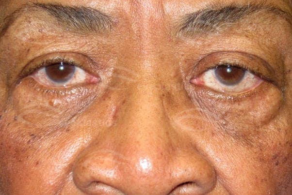
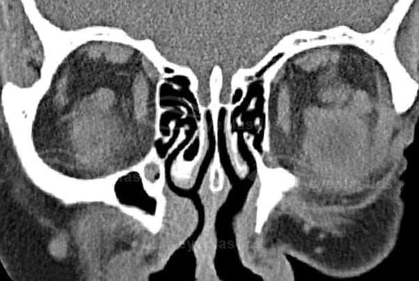
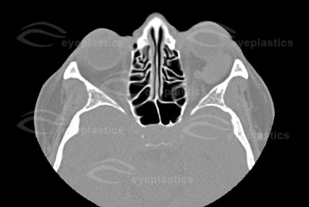
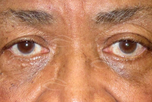
The Most Common Benign Orbital Tumor of Adults
A cavernous hemangioma — formally a cavernous venous malformation — is a benign, slow-growing vascular lesion composed of large, blood-filled channels within a well-defined fibrous capsule. It is the most common benign orbital tumor in adults, typically diagnosed in the 4th–5th decade (ages 30–60) and more often in women. Most arise within the muscle cone directly behind the eye, which is why their hallmark is a slow, painless, forward displacement of the globe.
Symptoms & Signs
- Gradual painless proptosis — the eye is slowly pushed forward over months to years; old photographs often reveal how long the change has been developing
- Hyperopic shift / blurred vision — pressure on the back of the eye shortens its focusing length and can cause retinal striae (choroidal folds)
- Double vision — from mass effect on the extraocular muscles in larger lesions
- Optic nerve compression — lesions at the orbital apex may affect vision or color vision even when small — the main reason apex lesions are managed differently
- Many are asymptomatic — found incidentally on a CT or MRI obtained for another reason
Imaging & Diagnosis
The diagnosis is usually made confidently on imaging without a biopsy. CT shows a well-circumscribed, round or oval intraconal mass. MRI is characteristic: the lesion is isointense to muscle on T1, brightly hyperintense on T2, and shows progressive “filling-in” enhancement on dynamic contrast sequences — contrast puddles slowly through the cavernous channels, a pattern that distinguishes it from other orbital tumors such as schwannoma or lymphangioma.
Treatment
- Observation — appropriate for small, asymptomatic, incidentally-found lesions: periodic eye exams, visual fields, and surveillance imaging
- Surgical excision — indicated for progressive proptosis, visual change, diplopia, or optic-nerve compression. Because the tumor is encapsulated and does not infiltrate, it can usually be dissected free and removed intact through an orbitotomy — lateral, anterior, or transconjunctival depending on its position — and complete excision is curative
- Apex lesions — lesions wedged at the orbital apex carry higher surgical risk to the optic nerve; management is individualized and may favor observation or staged approaches
Capillary vs. Cavernous — Same Word, Different Diseases
| Capillary (infantile) hemangioma | Cavernous hemangioma | |
|---|---|---|
| Who | Infants — appears in the first weeks of life | Adults — usually ages 30–60, women more often |
| Behavior | Grows rapidly, then shrinks away on its own (most gone by age 9) | Grows slowly and persists — never involutes |
| Location | Eyelid and front of the orbit, often a visible red lesion | Deep in the orbit behind the eye — usually nothing visible externally |
| Main risk | Amblyopia in the developing visual system | Proptosis and optic-nerve compression |
| Treatment | Observation; propranolol if vision threatened | Observation; surgical excision if symptomatic (curative) |
See the companion page: Capillary Hemangioma (infantile).
Ready to discuss Cavernous Hemangioma (Cavernous Venous Malformation)?
Schedule a consultation with Tamara R. Fountain, MD to learn if this procedure is right for you.